Challenges In Surgery
Here we present some of the challenges addressed by Ultravision in laparoscopic and open surgery.
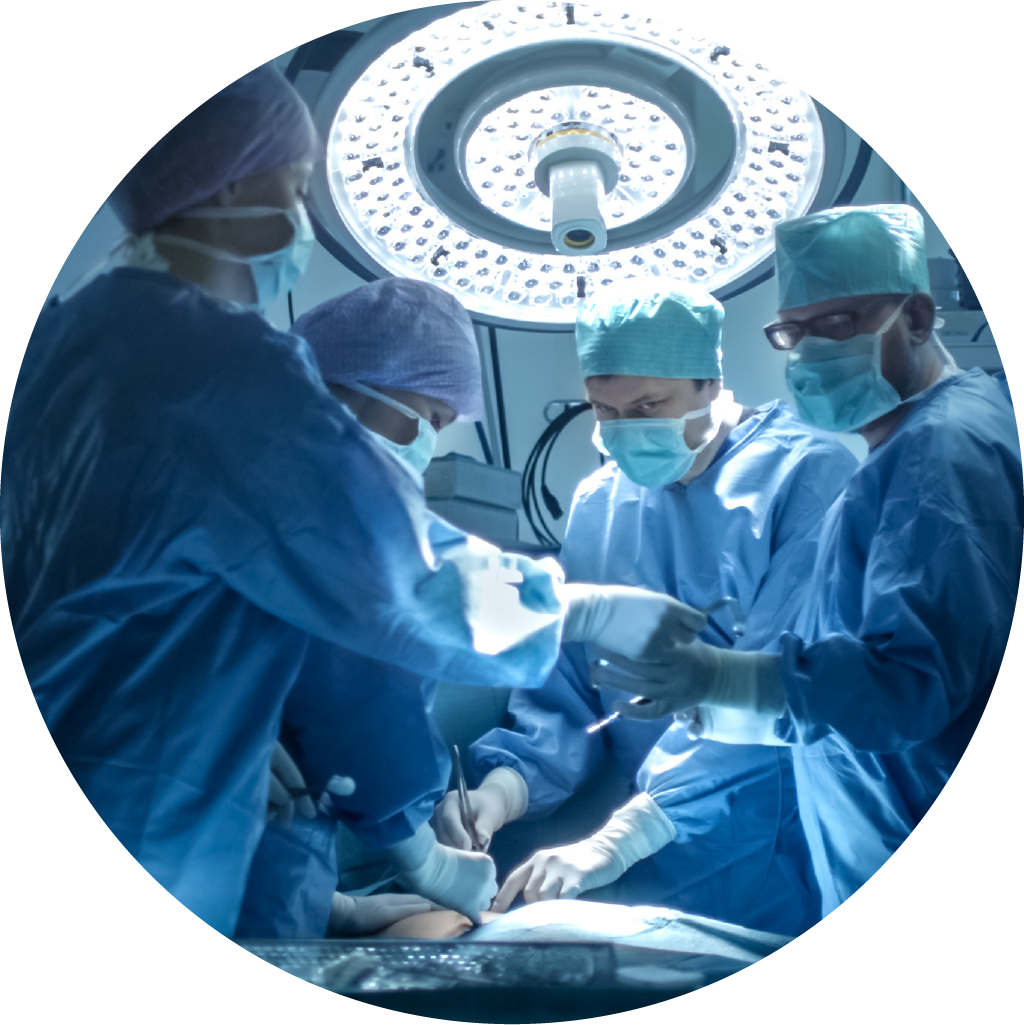
The perils of surgical
smoke in the OR and how
Ultravision overcomes the
limitations of other solutions
Here we summarise what is known on the subject of surgical smoke and how manufacturers have sought to deal with it. We also uncover the limitations of other systems and how Ultravision is advancing new protocols.
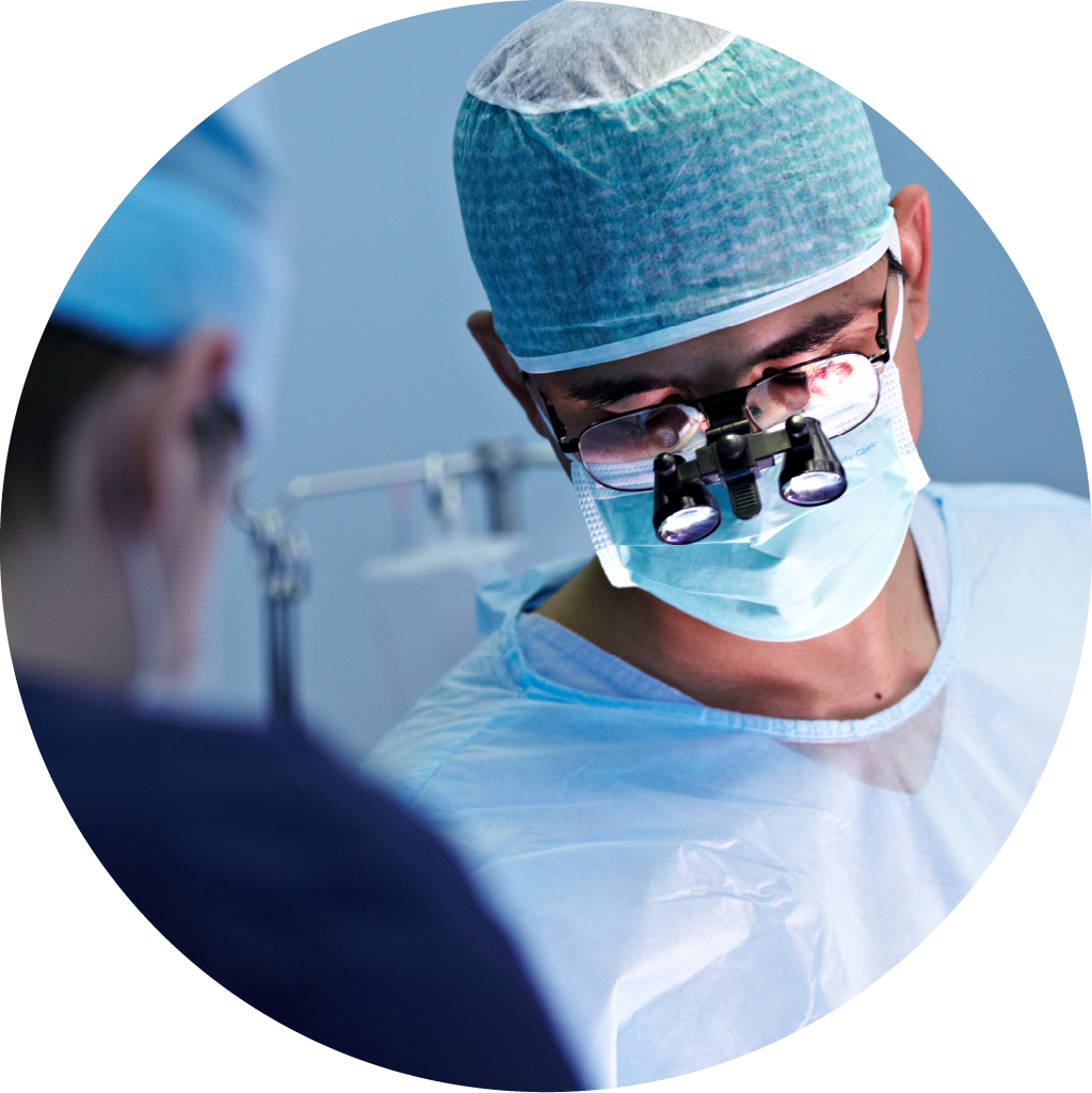
The importance of
visualization in
laparoscopic surgery
The evolution of electrosurgery has created new challenges to maintaining a clear line of sight for surgeons.
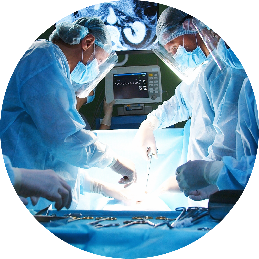
Controlling CO2 levels in
laparoscopic surgery
A review of how the technology that Ultravision employs, enables one of the key elements for ‘best practice’ laparoscopic surgery of establishing pneumoperitoneum.
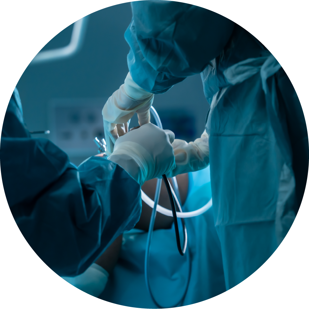
The importance of OR
efficiency
The pressure on healthcare providers to work more efficiently has never been greater. Here we explore how they manage their assets so that they can meet the ever-increasing demand for higher quality and more cost-effective provision of services.